StuDyiNG the CeLL
What are you made of ?
You could say atoms or elements. That would be right, but rocks & student desks & pizza boxes are made of atoms and elements too. What makes you different from those things ?
You could say organic compounds. That would be a better response I guess. Organic compounds come from living things. That answer would distinguish us from rocks & desks & pizza boxes. But other things are composed of organic compounds too; things like sugar and cow poop.
So I guess a better question is : what are you made of that makes you alive ?
A pretty big question, kind of philosophical. Since I'm a science teacher and not a philosopher, we'll just focus on the scientific units that make up each & every living thing on this planet : CELLS.
cell \'sel\ n 1 : the basic unit of structure & function in living things
In just a second we'll
review some of the dudes who were among the first to study cells.
In order for any of these guys to make any observations or discoveries,
there had to have been certain technological tools available to them. The
biggest of these tools was certainly the compound light microscope.
An understanding of the microscope is "a must" in any biology course.
I have dedicated
a page to the microscope in my "LAB REVIEW" Pages. To look at that
now, click here.
For now, here is a quick
exercise to review some important tools & techniques related to cell
study. Match the tool or technique with its description.
|
For Cell Study |
|
electron microscope microdissection apparatus phase-contrast microscope simple microscope staining stereomicroscope ultracentrifuge |
1. microscope
composed of one lens
2. microscope
that creates an image using two lenses
3. adding
a chemical that makes certain cell structures easier to see, usually kills
the cells
4. a high
resolution microscope used to study living cells
5. microscope
that provides images of the greatest magnification & resolution
6. microscope
with two oculars, usually used during dissections to observe relatively
large structures in more detail
7. small
tools used to remove or transplant cell organelles
8. machine
that can be used to separate cell organelles according to their densities
After you've done your best, check your answers <herE>.
OK, let's continue our review/study of the cell by reviewing the guys who first studied the cell. Number from 1-7 on a piece of paper & match each dude with his claim to fame.
![]()
|
Robert Hooke (1665) |
Matthias Schleiden (1838) |
Johannes Purkinje (1839) Rudolf Virchow (1858) |
1) first to use the term "protoplasm"Think you got 'em all right ? Click <here> for the answers.
2) while studying cork, he was the first to use the term "cell"
3) stated that all new cells come from other living cells
4) studied many microscopic organisms using a strong simple microscope
5) stated that all plants are composed of cells
6) stated that all animals are composed of cells
7) "discovered" the nucleus
THE CELL THEORY :
1. all living things are made up of cells &
the products of those cells
2. all cells carry out their own life functions
3. new cells come from other living cells
Exceptions to the
Cell Theory :
1. Viruses - are they alive
?
According
to the Cell Theory we have to say "no" because a virus is not a cell.
Viruses are made of two chemicals, protein & nucleic acid, but have
no membranes, nucleus, or protoplasm. They appear to be alive when they
reproduce after infecting a host cell.
2. Mitochondria &
chloroplasts.
These cell
organelles (small structures inside the cell) have their own genetic material
& reproduce independently from the rest of the cell.
3. Where did the first
cell come from ?
According
to statement #3 of the cell theory, all cells come from other living cells.
So how did the first cell ever appear ? It's the old "chicken &
egg" dilemma. We will investigate this question (& its possible
answer) in more detail during the Evolution Unit.
CELL STRUCTURE & ORGANELLES
ORGANELLE \or-ga-'nel\ n : 1. smaller structures within a cell, each organelle has a certain function.
PROTOPLASM \pro-to-paz-'um\ n : 1. general term for the living material in a cell
YOU HAVE TWO MISSIONS RELATED TO THIS SECTION :All cells have a cell membrane, cytoplasm and genetic material (DNA). From there things can vary in terms of the other organelles present in the cell, the shape of the cell, or the function of the cell.
Mission #1 : be able to identify all cell organelles in a diagram or picture
Mission #2 : be able to describe the function of each cell organelle
In this course we concern ourselves mostly with the differences between prokaryotic cells & eukaryotic cells, and between animal cells & plant cells.
The prokaryotic-eukaryotic difference
is easy; prokaryotic cells do not have a nucleus & eukaryotic cells
do. Remember that all prokaryotic organisms are classified in the Moneran
Kingdom. The organisms in the other four Kingdoms have eukaryotic cells.
(If you want to review the 5 Kingdoms,
check out my "5 Kingdoms Page".)
The animal-plant cell differences aren't too bad either. Basically, some organelles are found in plant cells and not animal cells & vice versa. More on that in a minute.
First ......
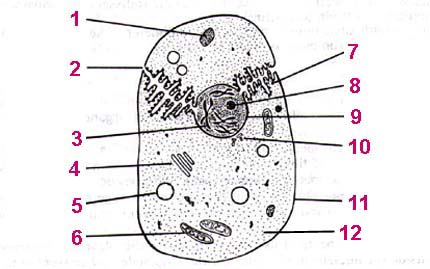
LET'S LABEL SOME ORGANELLES !!!
A little note : cell diagrams tend to be either overly complicated (fancy 3-D graphics) or too simplified (bare minimum "know this" stuff). Diagrams like the ones here have been seen in Regents Review Books & tests so I figure they are the most practical for us to use.

|
|
|
centrioles chromosomes (DNA) endoplasmic reticulum golgi body lysosome mitochondria nucleolus nucleus plasma membrane ribosome vacuole |
Check here
to click your work ---
I mean, click here
to check your work.
Before we label a plant cell, let's take a
sTudY BReaK !!!
OK, time to label a typical
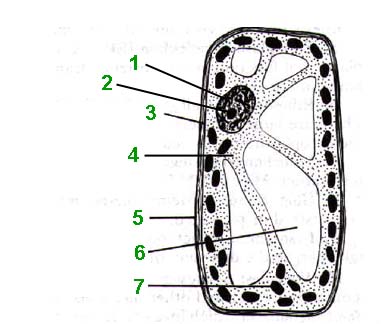
plant cell (notice the green?). Usually, plant cell diagrams focus on structures
that distinguish plant cells from animal cells. So some of the organelles
that animal & plant cells have in common (like ribosomes, golgi bodies,
endoplasmic reticulum) get left out all together. Label the diagram
below & see what I mean ...
PLANT CELL

|
|
|
chloroplast cytoplasm nucleolus nucleus plasma membrane vacuole (large) |
Take a look at the
answers.
PLANT CELLS vs ANIMAL CELLS
If you can label diagrams of a plant or animal cell, then you pretty much know what the differences are between them.This table summarizes the differences :
|
|
|
|
|
|
|
|
|
|
|
|
|
|
|
|
|
|
|
|
ORGANELLES & THEIR FUNCTIONS
Now that you know what
each organelle looks like, it's time to get the functions of each organelle
to stick to your brain somewhere. Choose an organelle from the word
bank for each description in #1-15.
|
|
|
cell wall chloroplast centrioles centrosome cytoplasm endoplasmic reticulum golgi apparatus lysosome mitochondria nuclear membrane nucleolus nucleus ribosomes vacuole |
1. liquid inside the cell, mostly water
2. made of lipids & proteins, it is the boundary of the cell; it controls what substances enter or leave the cell
3. "control center of the cell" where genetic material (DNA) is found
4. nonliving border that surrounds plant cells, made of cellulose
5. very small organelles that are the sites of protein synthesis
6. system of tubes through the cytoplasm involved in transporting materials
7. a flat stack of tubes involved in "packaging" materials that will exit the cell
8. site of cellular respiration (where energy is released from nutrients)
9. storage sac for water or other materials
10. controls what enters or exits the nucleus
11. dark round structure within the nucleus that produces ribosomes
12. specialized vacuole that stores digestive enzymes
13. structure in animal cells involved in cell division
14. spherical structure that contains the centrioles
15. site of photosynthesis in plant cells
OK, let's have even more fun & check your answers !
Um, some last minute thoughts on organelles :

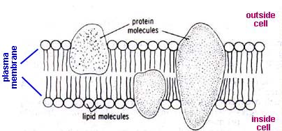
All of the molecules (lipids & proteins) "float" around, exchanging positions. This gives the membrane flexibility. The microscopic spaces between the molecules allow certain materials to enter or leave the cell. This is the primary function of the plasma membrane. The membrane is described as "selectively permeable", meaning it selects what substances enter or leave. In other words, not all substances are allowed to pass through the membrane.
Now that you know everything
there is to know about cells, may I suggest you brush up on your understanding
of the microscope ?
Check out my Microscope
Page.
StuDyiNG
the CeLL - ANSWER
PAGES
Answers to "Tool & Techniques
for Cell Study" :
1. simple microscope
2. compound microscope
3. staining
4. phase-contrast microscope
5. electron microscope
6. stereomicroscope
7. microdissection apparatus
8. ultracentrifuge
<-- back
Answers to "Cell Scientists
Matching" :
1) J. Purkinje
2) R. Hooke
3) R. Virchow
4) A. von Leeuwenhoek
5) M. Schleiden
6) M. Schwann (has an "a" in last
name - animal cells)
7) R. Brown
<--back
Answers to "Animal Cell Diagram" :

1. lysosome
2. endoplasmic reticulum
3. chromosome (DNA)
4. golgi body (apparatus)
5. vacuole
6. mitochondria
7. ribosome
8. nucleolus
9. nucleus (nuclear membrane would also be OK)
10. centrioles
11. plasma membrane
12. cytoplasm
NOTES & TIPS :<--back--
#1 - generally, lysosomes are illustrated as "shaded-in" circles or ovals.
#2 - channels running through the cytoplasm
#3 - the genetic material of the cell. humans have 46 chromosomes in each body cell (except eggs & sperm). this cell is eukaryotic because its genetic material is inside a nucleus.
#4 - stacks of membranes (like pancakes)
#5 - an "empty" oval or circle
#6 - you can recognize the mitochondria by the zig-zag line that is usually drawn in them. these represent internal membranes where respiration reactions occur.
#7 - very small dots, sometimes on the endoplasmic reticulum, sometimes out floating in cytoplasm
#8 - drawn as a dark circle inside the nucleus
#9 - generally depicted as the largest "shaded-in" circle inside the cell
#10 - drawn in pairs, two cylinder-like structures at right angles to each other
#11 - the outer boundary
#12 - the watery fluid that everything else floats in
Answers to "Plant Cell Diagram":

1. nucleus
2. nucleolus
3. plasma membrane
4. cytoplasm
5. cell wall
6. vacuole
7. chloroplast
NOTES & TIPS :back to the plant cell diagram
#1 - looks like an animal cell nucleus --- usually the largest "shaded-in" circle
#2 - same as an animal cell --- dark circle inside the nucleus
#3 - the plasma membrane in a plant cell is just inside the cell wall
#4 - cytoplasm is cytoplasm, everything else floats around in it
#5 - outermost boundary in a plant cell, looks like a picture frame. it is nonliving, not very flexible, and made of a polysaccharide called cellulose
#6 - drawn as "empty" areas in the cell, usually very large in plant cells
#7 - small dark ovals. sometimes get confused with mitochondria --- remember that mitochondria have the zig-zag, chloroplats are usually shaded "solid"
ANSWERS to "Organelle Functions"
Matching :
1. cytoplasm
2. cell membrane
3. nucleus
4. cell wall
5. ribosomes
6. endoplasmic reticulum
7. golgi apparatus
8. mitochondria
9. vacuole
10. nuclear membrane
11. nucleolus
12. lysosome
13. centrioles
14. centrosome
15. chloroplast
<--
back
sTudYBReaK
!!!
