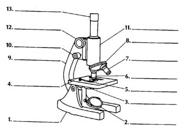
That gizmo pictured to the left is a BIG deal.
It literally opened up worlds of organisms and information to scientists.
It's importance in the history of medicine and our understanding of disease
should not be underestimated.
That gizmo is a compound light microscope.
For you, the biology student, it is perhaps
the most important tool for you to understand. By the time you are
done toying with these pages (& reading your text & paying attention
in class), you should be able to :
1. name all of its parts and describe the function of eachLet's get started .........
2. explain how to carry the thing, properly prepare a slide, & focus correctly
3. calculate total magnification
4. estimate the size of a specimen being observed
THE
PARTS
Match the names in the word
bank with the numbered parts in the picture.
|
base body tube coarse focus knob diaphragm fine focus knob high power objective lens light source low power objective lens nosepiece ocular (eyepiece) stage stage clips |

After you have jotted down the numbers & your answers, check your work here.
WHAT THE
PARTS DO
Now it's time to memorize
the function of each microscope part.
To help you practice, here's
a matching exercise.
|
base body tube coarse focus knob diaphragm fine focus knob high power objective lens light source low power objective lens nosepiece ocular (eyepiece) stage stage clips |
Want to see the answers ?
Important 'Scope Vocab :
magnification \mag-ne-fe-'ka-shen\ n 1. apparent enlargement of an object 2. the ratio of image size to actual sizeOK, well. There are a few other tidbits about the compound microscope you should remember :
A magnification of "100x" means that the image is 100 times bigger than the actual object.resolution \rez-e-loo-shen\ n 1. clarity, sharpness 2. the ability of a microscope to show two very close points separately
1. Why is called a "compound" light microscope ?
"Compound" just refers to the fact that there a two lenses magnifying the specimen at the same time, the ocular & one of the objective lenses.2. If two lenses are always magnifying the specimen
(see #1), how do you figure out the total magnification being used ?
You multiply the power of the ocular and the power of the objective being used.total mag. = ocular x objective For example, if the ocular is 10x and the low power objective is 20x, then the total magnification under low power is 10 x 20 = 200x.
Easy, ain't it ?3. How do you carry one of those things ?
With two hands, one holding the arm & the other under the base. Kinda like a football. (They're expensive, we don't want to drop 'em.)4. What about focussing ? How do you do that ?
Here's what I suggest. Once you have your slide in place on the stage, make sure the low power objective (the shortest objective lens) is in position & turn the coarse focus until the lens is at a position closest to the stage. Set the diaphragm to its largest opening (where it allows the most light through). Then, while looking through the ocular, begin to slowly turn the coarse focus. Turn slowly & watch carefully. When the specimen is focussed under low power, move the slide so that what you want to see is dead-center in your field of view, & then switch to a higher power objective. DO NOT touch the coarse focus again --- you will break something ! Once you are using a high power objective, focus using the fine focus knob ONLY. Be sure to center your specimen before switching to a higher power objective or it may disappear. More on that in a minute .....
WHAT YOU SEE
MICROSCOPIC MEASUREMENTS
Estimating Specimen
Size :
The area of the slide that you
see when you look through a microscope is called the "Field of View".
If you know how wide your field of view is, you can estimate the size of
things you see in the field of view. Figuring out the width of the field
of view is easy --- all you need is a thin metric ruler.
 By
carefully placing a thin metric ruler on the stage (where a slide would
usually go) and focussing under low power, we can measure the field
of view in millimeters. Through the microscope it would look something
like what you see here on the left. The total width of the field
of view in this example is less than 1.5 mm. A fair estimate would
be 1.3 or 1.4 mm.
(Relax, it's an estimate).
Now millimeters is a nice metric unit, but when we use a MICROscope we tend to use MICROmeters. To convert from millimeters to micrometers, move the decimal 3 places to the right. Our 1.3 mm estimate becomes 1300 micrometers.
Now we can get the ruler out of the way, prepare a slide, focus, and estimate the size of things we see ! (Exciting, ain't it ?)
For example, if something we were looking at took up half of the field of view, its size would be approximately 1/2 x 1300 micrometers = 650 micrometers. If something appeared to be 1/5 of the field of view, we would estimate its size to be 1/5 x 1300 = 260 micrometers.
Calculating Specimen
Size :
Because the high power objective
is so close to the stage, we can't measure the width of the field of view
under high power directly. The ruler just doesn't fit between the
objective & the stage. No problem. We can use the width
of the field of view under low power (which we measure using the steps
above) and the relationship between the low & high power magnifications
to mathematically calculate the width of the field of view under high power.
First of all memorize this :
When switching from low to high power, the area in the field of view gets smaller & darker. (You see a smaller area of the slide under high power.) This is why centering what you want to see prior to switching to high power is so important.The fraction of the area seen under high power is the same as the ratio of the low & high power magnifications.
Um, huh ?
For example : if the low power objective is 20x and the high power objective is 40x, then under high power we will see 20/40 or 1/2 of the area of the slide we saw under low power.
This is
something that requires some practice. So here ya go. Calculate the
answers to these examples on some paper & then click on "answers".
(You'll
learn more if you try it yourself first.)
Example
#1:
ocular power = 10x
low power objective = 20x
high power objective = 50xa) What is the highest magnification you could get using this microscope ?
b) If the diameter of the low power field is 2 mm, what is the diameter of the high power field of view in mm? in micrometers ?
c) If 10 cells can fit end to end in the low power field of view, how many of those cells would you see under high power ?
ocular power = 10x
low power objective = 10x
high power objective = 40x
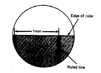
The diagram shows the edge of a millimeter ruler viewed under the microscope with the lenses listed above. The field shown is the low power field of view.
a) What is the approximate width of the field of view in micrometers ?
b) What would be the width of the field of view under high power ?
c) If 5 cells fit across the high power field of view, what is the approximate size of each cell ?
ocular = 10x
low power objective = 20x
high power objective = 40x
The picture shows the low power field of view for the microscope with the lenses listed above.
a) What is the approximate size of the cell in micrometers ?
b) What would be the high power field of view ?
c) How many cells like the one in the picture could fit in the high power field of view ?
Back to Biology Topics Outline

![]()
Answers to THE
PARTS :
1) base
2) light source 3) diaphragm 4) stage
5) stage clips
6) low
power objective lens 7) high power objective lens
8) nosepiece 9) arm 10) fine focus knob 11)
body tube 12) coarse focus knob 13) ocular (eyepiece)
<-- back to 'WHAT
THE PARTS DO'
Answers to 'WHAT YOU
SEE' :
ANSWER
to Example #1:
ocular power = 10xANSWER to example #2:
low power objective = 20x
high power objective = 50xa) What is the highest magnification you could get using this microscope ?500x
Ocular x high power = 10 x 50 = 500. (We can only use 2 lenses at a time, not all three.)
b) If the diameter of the low power field is 2 mm, what is the diameter of the high power field of view in mm ? .8 mm
The ratio of low to high power is 20/50. So at high power you will see 2/5 of the low power field of view (2 mm). 2/5 x 2 = 4/5 = .8 mm
in micrometers ? 800 micrometers
To convert mm to micrometers, move the decimal 3 places to the right (multiply by 1000). .8 mm x 1000 = 800 micrometers
d) If 10 cells can fit end to end in the low power field of view, how many of those cells would you see under high power ?4 cells.
We can answer this question the same way we go about "b" above. At high power we would see 2/5 of the low field. 2/5 x 10 cells = 4 cells would be seen under high power.
ocular power = 10x
low power objective = 10x
high power objective = 40x
The diagram shows the edge of a millimeter ruler viewed under the microscope with the lenses listed above. The field shown is the low power field of view.
a) What is the approximate width of the field of view in micrometers ? 3500 - 3800 micrometers
Each white space is 1 mm. We can see approximately 3 1/2 (or so) white spaces. That is equivalent to 3.5 mm, which converts to 3500 micrometers. Any answer in the range above would be OK.
b) What would be the width of the field of view under high power ?
875 micrometers
The ratio of low to high power for this microscope is 10/40 or 1/4. So, under high power we will see 1/4 of the low power field of view. 1/4 x 3500 micrometers (from "a" above) = 875 micrometers.
c) If 5 cells fit across the high power field of view, what is the approximate size of each cell ?
175 micrometers
If 5 cells fit in the high power field of view (which we determined is 875 micrometers in "b"), then the size of 1 cell = 875/5 = 175 micrometers.
ocular = 10x
low power objective = 20x
high power objective = 40x
The picture shows the low power field of view for the microscope with the lenses listed above.
a) What is the approximate size of the cell in micrometers ?
500 micrometers
First, we have to visualize how many of those cells could fit across the field --- about 4. So 2 mm (the width of the field) / 4 = .5 mm, which converts to 500 micrometers.
b) What would be the high power field of view ?
1000 micrometers
The ratio of low to high power for this scope is 20/40, or 1/2. So we will see 1/2 of the low power field under high power. 1/2 x 2 mm = 1mm, which converts to 1000 micrometers.
c) How many cells like the one in the picture could fit in the high power field of view ?
2 cells
Again the ratio of low to high power is 20/40, or 1/2. If we can see 4 cells across the low field of view we will see 1/2 as many in the high field of view. 1/2 x 4 = 2 cells.